上海金畔生物科技有限公司为生命科学和医药研发人员提供生物活性分子抑制剂、激动剂、特异性抑制剂、化合物库、重组蛋白,专注于信号通路和疾病研究领域。
Empesertib (Synonyms: BAY 1161909) 纯度: ≥98.0%
Empesertib (BAY 1161909) 是一种有效的 Mps1 抑制剂,IC50 值 <1 nM。

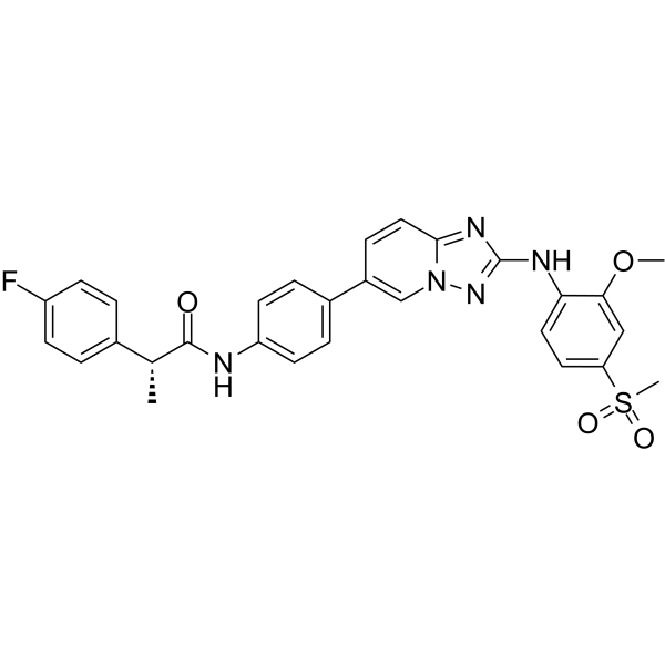
Empesertib Chemical Structure
CAS No. : 1443763-60-7
| 规格 | 价格 | 是否有货 | 数量 |
|---|---|---|---|
| 10 mM * 1 mL in DMSO | ¥4309 | In-stock | |
| 2 mg | ¥2333 | In-stock | |
| 5 mg | ¥3500 | In-stock | |
| 10 mg | ¥5000 | In-stock | |
| 50 mg | 询价 | ||
| 100 mg | 询价 |
* Please select Quantity before adding items.
Empesertib 相关产品
•相关化合物库:
- Clinical Compound Library Plus
- Bioactive Compound Library Plus
- Cell Cycle/DNA Damage Compound Library
- Kinase Inhibitor Library
- Anti-Cancer Compound Library
- Clinical Compound Library
- Anti-Aging Compound Library
- Cytoskeleton Compound Library
| 生物活性 |
Empesertib (BAY 1161909) is a potent Mps1 inhibitor, with an IC50 of < 1 nM. |
||||||||||||||||
|---|---|---|---|---|---|---|---|---|---|---|---|---|---|---|---|---|---|
| IC50 & Target |
|
||||||||||||||||
| 体外研究 (In Vitro) |
BAY 1161909 is a potent Mps1 inhibitor, with an IC50 of < 1 nM. BAY 1161909 also shows anti-tumor effect, suppressing the proliferation of Hela cells, with an IC50 of < 400 nM[1]. 上海金畔生物科技有限公司 has not independently confirmed the accuracy of these methods. They are for reference only. |
||||||||||||||||
| Clinical Trial |
|
||||||||||||||||
| 分子量 |
559.61 |
||||||||||||||||
| Formula |
C29H26FN5O4S |
||||||||||||||||
| CAS 号 |
1443763-60-7 |
||||||||||||||||
| 运输条件 |
Room temperature in continental US; may vary elsewhere. |
||||||||||||||||
| 储存方式 |
|
||||||||||||||||
| 溶解性数据 |
In Vitro:
DMSO : ≥ 35 mg/mL (62.54 mM) * “≥” means soluble, but saturation unknown. 配制储备液
*
请根据产品在不同溶剂中的溶解度选择合适的溶剂配制储备液;一旦配成溶液,请分装保存,避免反复冻融造成的产品失效。 In Vivo:
请根据您的实验动物和给药方式选择适当的溶解方案。以下溶解方案都请先按照 In Vitro 方式配制澄清的储备液,再依次添加助溶剂: ——为保证实验结果的可靠性,澄清的储备液可以根据储存条件,适当保存;体内实验的工作液,建议您现用现配,当天使用; 以下溶剂前显示的百
|
||||||||||||||||
| 参考文献 |
|
| Kinase Assay [1] |
N-terminally GST-tagged human full length recombinant Mps-1 kinase is used. As substrate for the kinase reaction a biotinylated peptide of the amino-acid sequence PWDPDDADITEILG is used. For the assay 50 nL of a 100-fold concentrated solution of the test compounds (BAY 1161909, etc.) in DMSO is pipetted into a black low volume 384 well microtiter plate, 2 μL of a solution of Mps-1 in assay buffer [0.1 mM sodium-ortho-vanadate, 10 mM MgCl2, 2 mM DTT, 25 mM Hepes pH 7.7, 0.05% BSA, 0.001 % Pluronic F-127] are added and the mixture is incubated for 15 min at 22°C to allow pre-binding of the test compounds to Mps-1 before the start of the kinase reaction. Then the kinase reaction is started by the addition of 3 μL of a solution of 16.7 adenosine-tri-phosphate and peptide substrate in assay buffer and the resulting mixture is incubated for a reaction time of 60 min at 22°C. The concentration of Mps-1 in the assay is adjusted to the activity of the enzyme lot and is chosen appropriate to have the assay in the linear range, typical enzyme concentrations are in the range of about 1 nM (final cone, in the 5 μL assay volume). The reaction is stopped by the addition of 3 μL of a solution of HTRF detection reagents (100 mM Hepes pH 7.4, 0.1 % BSA, 40 mM EDTA, 140 nM Streptavidin-XLent, 1 .5 nM anti-phospho(Ser/Thr)-Europium-antibody[1]. 上海金畔生物科技有限公司 has not independently confirmed the accuracy of these methods. They are for reference only. |
|---|---|
| Cell Assay [1] |
Cultivated tumor cells are plated at a density of 5000 cells/ well (MCF7, DU145, HeLa-MaTu-ADR), 3000 cells/well (NCI-H460, HeLa-MaTu, HeLa), or 1000 cells/well (B16F10) in a 96-well multititer plate in 200 μL of their respective growth medium supplemented 10% fetal calf serum. After 24 hours, the cells of one plate (zero-point plate) are stained with crystal violet, while the medium of the other plates is replaced by fresh culture medium (200 μL), to which the test substances (BAY 1161909, etc.) are added in various concentrations (0 μM, as well as in the range of 0.01-30 μM; the final concentration of the solvent DMSO is 0.5%). The cells are incubated for 4 days in the presence of test substances. Cell proliferation is determined by staining the cells with crystal violet: the cells are fixed by adding 20 μL/measuring point of an 11% glutaric aldehyde solution for 15 minutes at room temperature. After three washing cycles of the fixed cells with water, the plates are dried at room temperature. The cells are stained by adding 100 μL/measuring point of a 0.1% crystal violet solution (pH 3.0). After three washing cycles of the stained cells with water, the plates are dried at room temperature. The dye is dissolved by adding 100 μL/measuring point of a 10% acetic acid solution. The IC50 values are determined by means of a 4 parameter fit. The compounds A1 (BAY 1161909), A2, A3, A4 and A5 are characterized by an IC50 determined in a HeLa-MaTu-ADR cell proliferation assay[1]. 上海金畔生物科技有限公司 has not independently confirmed the accuracy of these methods. They are for reference only. |
| 参考文献 |
|
所有产品仅用作科学研究或药证申报,我们不为任何个人用途提供产品和服务
