上海金畔生物科技有限公司为生命科学和医药研发人员提供生物活性分子抑制剂、激动剂、特异性抑制剂、化合物库、重组蛋白,专注于信号通路和疾病研究领域。
Y-33075 (Synonyms: Y 39983) 纯度: 99.19%
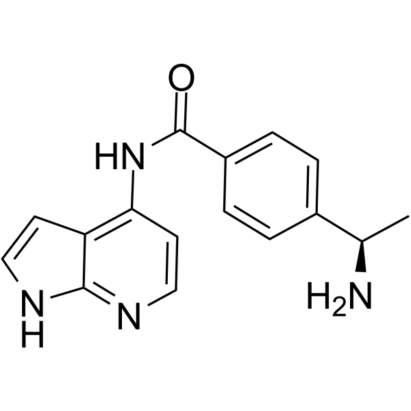
Y-33075 是一种有效的 ROCK 抑制剂,源于 Y-27632,但活性更强,IC50 值 3.6 nM。

Y-33075 Chemical Structure
CAS No. : 199433-58-4
| 规格 | 价格 | 是否有货 | 数量 |
|---|---|---|---|
| Free Sample (0.1-0.5 mg) | Apply now | ||
| 10 mM * 1 mL in DMSO | ¥2814 | In-stock | |
| 10 mg | ¥2558 | In-stock | |
| 50 mg | ¥9207 | In-stock | |
| 100 mg | 询价 | ||
| 200 mg | 询价 |
* Please select Quantity before adding items.
Y-33075 相关产品
•相关化合物库:
- Bioactive Compound Library Plus
- Cell Cycle/DNA Damage Compound Library
- Kinase Inhibitor Library
- Stem Cell Signaling Compound Library
- TGF-beta/Smad Compound Library
- Anti-Cancer Compound Library
- Anti-Aging Compound Library
- Reprogramming Compound Library
- Cytoskeleton Compound Library
| 生物活性 |
Y-33075 is a selective ROCK inhibitor derived from Y-27632, and is more potent than Y-27632, with an IC50 of 3.6 nM. |
||||||||||||||||
|---|---|---|---|---|---|---|---|---|---|---|---|---|---|---|---|---|---|
| IC50 & Target[1] |
|
||||||||||||||||
| 体外研究 (In Vitro) |
Y-33075 (Y-39983) is a potent ROCK inhibitor, with an IC50 of 3.6 nM. Y-33075 also inhibits PKC and CaMKII more potently than Y-27632, and the IC50s of Y-27632 and Y-33075 for PKC are 9.0 μM and 0.42 μM, respectively, whereas the IC50s of Y-27632 and Y-33075 for CaMKII are 26 μM and 0.81 μM, respectively. The IC50s of Y-27632 and Y-33075 for PKC is 82 and 117 times those for ROCK, respectively, whereas the IC50s of Y-27632 and Y-33075 for CaMKII is 236 and 225 times those for ROCK, respectively[1]. Y-33075 (Y-39983, 10 μM) extends neurites in the retinal ganglion cells (RGCs) compared with those in RGCs treated without Y-39983[2]. Y-33075 (Y-39983, 1 μM) inhibits the contraction of rabbit ciliary artery segments evoked by histamine in Ca2+-free solutions. Y-33075 (10 μM) shows no effect on the [Ca2+]i increase with the high-potassium (high-K) solution[3]. 上海金畔生物科技有限公司 has not independently confirmed the accuracy of these methods. They are for reference only. |
||||||||||||||||
| 体内研究 (In Vivo) |
In rabbits, Y-39983 (≥0.01%) significantly lowers intraocular pressure (IOP) at 2 hours after topical administration. In monkeys, Y-39983 (0.05%)-treated eyes show significant reduction of IOP between 2 and 7 hours after topical administration[1]. Y-39983 (100 μM) increases the regenerating axons of retinal ganglion cells (RGCs) in the eyes of the rats[2]. 上海金畔生物科技有限公司 has not independently confirmed the accuracy of these methods. They are for reference only. |
||||||||||||||||
| 分子量 |
280.32 |
||||||||||||||||
| Formula |
C16H16N4O |
||||||||||||||||
| CAS 号 |
199433-58-4 |
||||||||||||||||
| 运输条件 |
Room temperature in continental US; may vary elsewhere. |
||||||||||||||||
| 储存方式 |
|
||||||||||||||||
| 溶解性数据 |
In Vitro:
DMSO : 50 mg/mL (178.37 mM; Need ultrasonic and warming) H2O : 1 mg/mL (3.57 mM; ultrasonic and heat to 60°C) 配制储备液
*
请根据产品在不同溶剂中的溶解度选择合适的溶剂配制储备液;一旦配成溶液,请分装保存,避免反复冻融造成的产品失效。 In Vivo:
请根据您的实验动物和给药方式选择适当的溶解方案。以下溶解方案都请先按照 In Vitro 方式配制澄清的储备液,再依次添加助溶剂: ——为保证实验结果的可靠性,澄清的储备液可以根据储存条件,适当保存;体内实验的工作液,建议您现用现配,当天使用; 以下溶剂前显示的百
|
||||||||||||||||
| 参考文献 |
|
| Kinase Assay [1] |
Recombinant ROCK (ROK α/ROCK II), purified protein kinase C (PKC: mixture of α, β, γ isoforms), and recombinant calmodulin-dependent protein kinase II (CaMK II) are used in the assay. ROCK (0.2 U/mL) is incubated with 1 μM [γ-32P] ATP and 10 μg/mL histone as substrates in the absence or presence of various concentrations of Y-27632, Y-33075, or staurosporine at room temperature for 20 minutes in 20 mM MOPS (3-(N-morpholino)propanesulfonic acid) buffer (pH 7.2) containing 0.1 mg/mL bovine serum albumin (BSA), 5 mM dithiothreitol [DTT], 10 mM β-glycerophosphate, 50 μM Na3VO4, and 10 mM MgCl2 in a total volume of 100 μL. PKC (10 ng/mL) is incubated with 1 μM [γ-32P] ATP and 20 μM PKC substrate in the absence or presence of various concentrations of Y-27632, Y-33075, or staurosporine at room temperature for 30 minutes in 20 mM MOPS buffer (pH 7.5) containing 0.1 mg/mL BSA, 10 mM DTT, 10 mM β-glycerophosphate, 50 μM Na3VO4, 2 mM CaCl2, 20 μg/mL phosphatidyl-l-serine, and 10 mM MgCl2 in a total volume of 100 μL. CaMK II (125 U/mL) is incubated with 1 μM [γ-32P] ATP, 10 μM calmodulin, and 20 μM CaMK II substrate, in the absence or presence of various concentrations of Y-27632, Y-33075, or staurosporine at room temperature for 30 minutes in 20 mM MOPS buffer (pH 7.5) containing 0.2 mg/mL BSA, 0.5 mM DTT, 0.1 mM β-glycerophosphate, 50 μM Na3VO4, 1 mM CaCl2, and 5 mM MgCl2 in a total volume of 100 μL. Incubation is terminated by the addition of 100 μL of 0.7% phosphoric acid. A 160 μL portion of the mixture is transferred to Multiscreen-PH plate. A positively charged phosphocellulose filter absorbs the substrate that binds 32P. The filter is washed with 300 μL of 0.5% phosphoric acid and then twice with purified water and then dried. The radioactivity of the dried filter is measured with a liquid scintillation counter. Results are presented as 50% inhibitory concentrations and 95% confidence intervals (CIs)[1]. 上海金畔生物科技有限公司 has not independently confirmed the accuracy of these methods. They are for reference only. |
|---|---|
| Cell Assay [2] |
In brief, retinal cell suspensions are obtained from dissected retinas of Wistar rats by papain treatment. Retinal ganglion cells (RGCs) are purified by the panning method using anti-rat CD11 antibody for removal of microglia cells and anti-rat Thy-1 antibody for isolation of ganglion cells. The purified RGCs (5000 cells/plate) are seeded into 24-well plates coated by 50 μg/mL of poly-l-lysine and 2 μg/mL of merosin, and are cultured in serum-free neurobasal medium supplemented with 2% B27 supplement, 50 ng/mL BDNF, 50 ng/mL CNTF, 5 μM forskolin, and 1 mM glutamine under a 95% air-5% CO2 atmosphere at 37°C. After completion of 24-hour cultivation, RGCs are cultured in medium with or without 10 μM Y-33075 as the final concentration for 24 hours and morphologically observed by phase-contrast microscopy. The concentration used is determined based on the effect of Y-33075 on trabecular meshwork contraction in vitro. Since this study is conducted in order to confirm whether Y-33075 has a potential of effect on axonal regeneration of RGCs, the effect is unquantitateively evaluated[2]. 上海金畔生物科技有限公司 has not independently confirmed the accuracy of these methods. They are for reference only. |
| Animal Administration [2] |
In brief, SD rats is anesthetized with an intraperitoneal injection of sodium pentobarbital (0.4 mg/kg body weight), and the optic nerve of one eye is transected 4 to 6 mm posterior to the eyeball, taking care to avoid injury to the ophthalmic artery. The anterior branch of the sciatic nerve is excised and sutured autologously to the optic nerve stump with nylon sutures. The other end of the graft is sutured to the temporalis muscle. A small piece (3 mm × 3 mm) of gelatin sponge soaked with 10 μM Y-33075 or saline as a control is implanted in the space behind the optic stump after optic nerve transection in intact animals. Five μL of 0.12 mM or 1.2 mM Y-33075 solution or saline is administered into the vitreous body to final concentrations of 10 μM or 100 μM, respectively. The concentrations of Y-33075 used is determined as 10 μM that is effective in the in vitro study on axonal regeneration of RGCs, and also as 100 μM in order to confirm the dose response of Y-33075. Six weeks after surgery, rats is anesthetized with an intraperitoneal injection of sodium pentobarbital (0.4 mg/kg body weight), and 4-Di-10ASP is embedded in the transplanted sciatic nerve to retrogradely label RGCs with axonal regeneration into the sciatic nerve. Three days after dye embedding, rats is euthanized and the eyes is enucleated for preparation of retinal flat-mounts. The posterior eyecup is then separated from the vitreous body and postfixed with 4% paraformaldehyde solution in phosphate buffer for around 1 hour at room temperature. Fluorescence micrographs of the labeled cells is imported using a fluorescence microscope connected to a computer. Labeled cells is counted using image analysis software. As a normal group, the subsequent procedure for retrograde labeling with 4-Di-10ASP is performed without grafting sciatic nerve and administering the test drug. Statistical analysis is performed using logarithmically transformed values due to differences in variance among the groups. The statistical significance of differences between the normal and saline groups and the saline and Y-33075 groups is examined by t-test (onesided) and William’s test (one-sided). Findings of p < 0.05 is considered significant[2]. 上海金畔生物科技有限公司 has not independently confirmed the accuracy of these methods. They are for reference only. |
| 参考文献 |
|
所有产品仅用作科学研究或药证申报,我们不为任何个人用途提供产品和服务
