上海金畔生物科技有限公司为生命科学和医药研发人员提供生物活性分子抑制剂、激动剂、特异性抑制剂、化合物库、重组蛋白,专注于信号通路和疾病研究领域。
BI-78D3 纯度: 99.49%
BI-78D3 是一种底物竞争性的 JNK 抑制剂,抑制 JNK 激酶活性,IC50 为 280 nM。

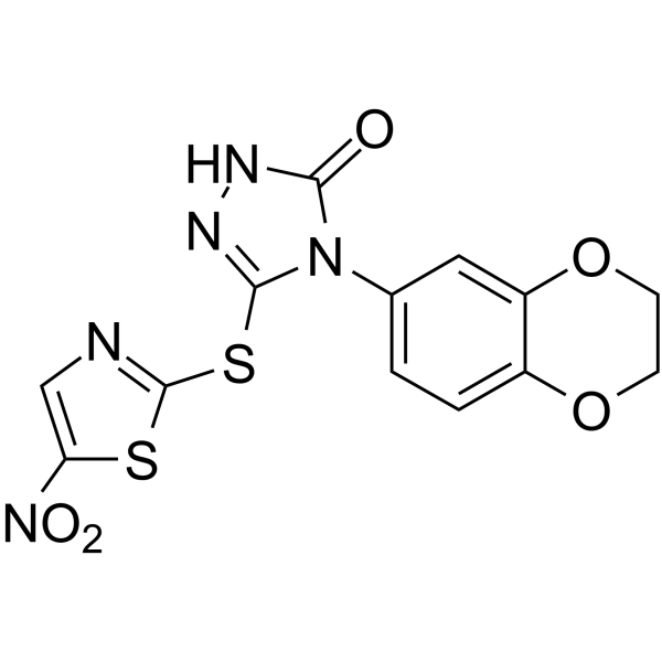
BI-78D3 Chemical Structure
CAS No. : 883065-90-5
| 规格 | 价格 | 是否有货 | 数量 |
|---|---|---|---|
| Free Sample (0.1-0.5 mg) | Apply now | ||
| 10 mM * 1 mL in DMSO | ¥968 | In-stock | |
| 5 mg | ¥880 | In-stock | |
| 10 mg | ¥1350 | In-stock | |
| 25 mg | ¥2850 | In-stock | |
| 50 mg | ¥4950 | In-stock | |
| 100 mg | ¥8100 | In-stock | |
| 200 mg | 询价 | ||
| 500 mg | 询价 |
* Please select Quantity before adding items.
BI-78D3 相关产品
•相关化合物库:
- Bioactive Compound Library Plus
- Immunology/Inflammation Compound Library
- Kinase Inhibitor Library
- MAPK Compound Library
- Anti-Cancer Compound Library
- Reprogramming Compound Library
- Diabetes Related Compound Library
- Oxygen Sensing Compound Library
- Anti-Parkinson’s Disease Compound Library
- Anti-Obesity Compound Library
- Transcription Factor Targeted Library
- Targeted Diversity Library
| 生物活性 |
BI-78D3 functions as a substrate competitive inhibitor of JNK, inhibit the JNK kinase activity (IC50=280 nM). |
||||||||||||||||
|---|---|---|---|---|---|---|---|---|---|---|---|---|---|---|---|---|---|
| IC50 & Target[1] |
|
||||||||||||||||
| 体外研究 (In Vitro) |
BI-78D3, dose-dependently inhibits the phosphorylation of JNK substrates both in vitro and in cell. BI-78D3 is able to compete with the D-domain of JIP1 (amino acids 153-163; pepJIP1) for JNK1 binding (IC50=500 nM). Using the same in vitro LanthaScreen kinase assay and the same ATF2 substrate, BI-78D3 is found to be 100-fold less active vs. p38α, a member of the MAPK family with high structural similarity to JNK, and completely inactive against mTOR and PI3-kinase (α-isoform), both unrelated protein kinases. Furthermore, Lineweaver-Burk analysis clearly indicates that BI-78D3 is competitive with ATF2 for binding to JNK1 with an apparent Ki value of 200 nM. In an attempt to profile the properties of BI-78D3 in the context of a complex cellular milieu, the cell-based LanthaScreen kinase assay is used. In this assay BI-78D3 is able to inhibit TNF-α stimulated phosphorylation of c-Jun in cell (EC50=12.4 μM)[1]. 上海金畔生物科技有限公司 has not independently confirmed the accuracy of these methods. They are for reference only. |
||||||||||||||||
| 体内研究 (In Vivo) |
The link between ConA-induced liver failure, TNF receptor signaling, and JNK function has been established by studies employing JNK1-/- and JNK2-/- mice. For this analysis, insulin insensitive mice are injected only once with 25 mg/kg BI-78D3, 30 min before insulin injection. The effect of insulin on blood glucose levels is then measured. BI-78D3 results in a statistically significant reduction in blood glucose levels as compared with the vehicle control. Thus, the ability of BI-78D3 to abrogate ConA-induced liver damage and restore insulin sensitivity is consistent with its proposed function as an effective JNK inhibitor. Liquid chromatography/mass spectrometry bio-availability analysis demonstrates that BI-78D3 has favorable microsome and plasma stability (T1/2=54 min)[1]. 上海金畔生物科技有限公司 has not independently confirmed the accuracy of these methods. They are for reference only. |
||||||||||||||||
| 分子量 |
379.37 |
||||||||||||||||
| Formula |
C13H9N5O5S2 |
||||||||||||||||
| CAS 号 |
883065-90-5 |
||||||||||||||||
| 运输条件 |
Room temperature in continental US; may vary elsewhere. |
||||||||||||||||
| 储存方式 |
|
||||||||||||||||
| 溶解性数据 |
In Vitro:
DMSO : 100 mg/mL (263.59 mM; Need ultrasonic) 配制储备液
*
请根据产品在不同溶剂中的溶解度选择合适的溶剂配制储备液;一旦配成溶液,请分装保存,避免反复冻融造成的产品失效。 In Vivo:
请根据您的实验动物和给药方式选择适当的溶解方案。以下溶解方案都请先按照 In Vitro 方式配制澄清的储备液,再依次添加助溶剂: ——为保证实验结果的可靠性,澄清的储备液可以根据储存条件,适当保存;体内实验的工作液,建议您现用现配,当天使用; 以下溶剂前显示的百
|
||||||||||||||||
| 参考文献 |
|
| Kinase Assay [1] |
The cell based kinase assays for c-Jun and ATF2 phosphorylation carry out by using the LanthaScreen c-Jun (1-79) HeLa and LanthaScreen ATF2 (19-106) A549 cell lines which stably express GFP-c-Jun 1-79 and GFP-ATF2 19-106, respectively. Phosphorylation is determined by measuring the time resolved FRET (TR-FRET) between a terbium labeled phospho-specific antibody and the GFP-fusion protein. The cells are plated in white tissue culture treated 384 well plates at a density of 10,000 cell per well in 32 μl assay medium (supplemented with 1% charcoal/dextran-treated FBS, 100 U/mL Penicillin and 100 μg/mL Streptomycin, 0.1 mM nonessential amino acids, 1 mM sodium pyruvate, 25 mM Hepes, pH 7.3, and lacking phenol red). After overnight incubation, cells are pretreated for 60 min with BI-78D3 (0.001, 0.01, 0.1, 1, 10, and 100 μM) followed by 30 min of stimulation with 2 ng/mL of TNF-α that stimulates both JNK and p38. The medium is then removed by aspiration and the cells are lysed by adding 20 μL of lysis buffer (20 mM Tris•HCl, pH 7.6, 5 mM EDTA, 1% Nonidet P-40 substitute, 5 mM NaF, 150 mM NaCl, and 1:100 protease and phosphatase inhibitor mix, SIGMA P8340 and P2850, respectively). The lysis buffer includs 2 nM of the terbium-labeled anti-pc-Jun (pSer73) or anti-pATF2 (pThr71) detection antibodies. After allowing the assay to equilibrate for 1 h at room temperature, TR-FRET emission ratios are determined on a BMG Pherastar fluorescence plate reader (excitation at 340 nm, emission 520 nm and 490 nm; 100 μs lag time, 200 μs integration time, emission ratio=Em520/Em 490)[1]. 上海金畔生物科技有限公司 has not independently confirmed the accuracy of these methods. They are for reference only. |
|---|---|
| Cell Assay [1] |
Mice[1] 上海金畔生物科技有限公司 has not independently confirmed the accuracy of these methods. They are for reference only. |
| 参考文献 |
|
所有产品仅用作科学研究或药证申报,我们不为任何个人用途提供产品和服务
