上海金畔生物科技有限公司为生命科学和医药研发人员提供生物活性分子抑制剂、激动剂、特异性抑制剂、化合物库、重组蛋白,专注于信号通路和疾病研究领域。
3CAI 纯度: 99.97%
3CAI 是一种有效的特异性 AKT1 和 AKT2 抑制剂。

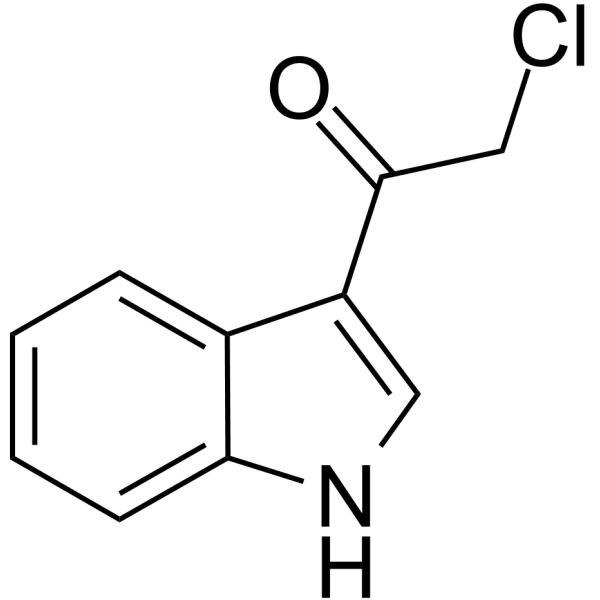
3CAI Chemical Structure
CAS No. : 28755-03-5
| 规格 | 价格 | 是否有货 | 数量 |
|---|---|---|---|
| Free Sample (0.1-0.5 mg) | Apply now | ||
| 10 mM * 1 mL in DMSO | ¥638 | In-stock | |
| 5 mg | ¥580 | In-stock | |
| 10 mg | ¥900 | In-stock | |
| 50 mg | ¥2600 | In-stock | |
| 100 mg | 询价 | ||
| 200 mg | 询价 |
* Please select Quantity before adding items.
3CAI 相关产品
•相关化合物库:
- Bioactive Compound Library Plus
- Kinase Inhibitor Library
- PI3K/Akt/mTOR Compound Library
- Stem Cell Signaling Compound Library
- Anti-Cancer Compound Library
- Autophagy Compound Library
- Anti-Aging Compound Library
- Differentiation Inducing Compound Library
- Oxygen Sensing Compound Library
- Glycolysis Compound Library
- Cytoskeleton Compound Library
- Glutamine Metabolism Compound Library
- Anti-Breast Cancer Compound Library
- Anti-Lung Cancer Compound Library
- Anti-Pancreatic Cancer Compound Library
- Anti-Blood Cancer Compound Library
- Anti-Cancer Metabolism Compound Library
- Anti-Obesity Compound Library
- Angiogenesis Related Compound Library
- Glucose Metabolism Compound Library
- Targeted Diversity Library
- Anti-Liver Cancer Compound Library
- Anti-Colorectal Cancer Compound Library
| 生物活性 |
3CAI is a potent and specific AKT1 and AKT2 inhibitor. |
||||||||||||||||
|---|---|---|---|---|---|---|---|---|---|---|---|---|---|---|---|---|---|
| IC50 & Target[1] |
|
||||||||||||||||
| 体外研究 (In Vitro) |
3CAI is a potential inhibitor of AKT. Based on these screening data, the effect of 3CAI on the kinase activities of AKT1, MEK1, JNK1, ERK1 and TOPK is tested using in vitro kinase assays. The results show that 3CAI (1 μM) suppresses only AKT1 kinase activity and the other kinases tested are not affected by 3CAI. 3CAI is a much more potent AKT1 inhibitor than PI3K (60% inhibition at 1 vs 10 μM, respectively). 3CAI substantially suppresses AKT1 activity as well as AKT2 activity in a dose dependent manner. 3CAI inhibits down-stream targets of AKT and induces apoptosis. AKT-mediated phosphorlyation site of mTOR (Ser2448) and GSK3β (Ser9) are substantially decreased by 3CAI in a time-dependent manner. Furthermore, pro-apoptotic marker proteins p53 and p21 are also upregulated by 3CAI after 12 or 24 h of treatment. HCT116 and HT29 colon cancer cells are seeded on 6 cm dishes in 1% FBS/McCoy’s 5A (HCT116) with 3CAI (4 μM), I3C or the AKT inhibitor and then incubated for 4 days. Results show that the number of apoptotic cells is significantly increased by 3CAI in HCT116 and HT29 colon cancer cells compared with untreated control cells[1]. 上海金畔生物科技有限公司 has not independently confirmed the accuracy of these methods. They are for reference only. |
||||||||||||||||
| 体内研究 (In Vivo) |
To examine the antitumor activity of 3CAI in vivo, HCT116 cancer cells are injected into the right flank of individual athymic nude mice. Mice are orally administered 3CAI at 20 or 30 mg/kg, I3C at 100 mg/kg, or vehicle 5 times a week for 21 days. Treatment of mice with 30 mg/kg of 3CAI significantly suppresses HCT116 tumor growth by 50% relative to the vehicle-treated group (p<0.05). Remarkably, mice seem to tolerate treatment with these doses of 3CAI without overt signs of toxicity or significant loss of body weight compared with vehicle-treated group. Expression of these AKT-target proteins is strongly suppressed by 30 mg/kg of 3CAI in tumor tissues[1]. 上海金畔生物科技有限公司 has not independently confirmed the accuracy of these methods. They are for reference only. |
||||||||||||||||
| 分子量 |
193.63 |
||||||||||||||||
| Formula |
C10H8ClNO |
||||||||||||||||
| CAS 号 |
28755-03-5 |
||||||||||||||||
| 运输条件 |
Room temperature in continental US; may vary elsewhere. |
||||||||||||||||
| 储存方式 |
|
||||||||||||||||
| 溶解性数据 |
In Vitro:
DMSO : 250 mg/mL (1291.12 mM; Need ultrasonic) 配制储备液
*
请根据产品在不同溶剂中的溶解度选择合适的溶剂配制储备液;一旦配成溶液,请分装保存,避免反复冻融造成的产品失效。 In Vivo:
请根据您的实验动物和给药方式选择适当的溶解方案。以下溶解方案都请先按照 In Vitro 方式配制澄清的储备液,再依次添加助溶剂: ——为保证实验结果的可靠性,澄清的储备液可以根据储存条件,适当保存;体内实验的工作液,建议您现用现配,当天使用; 以下溶剂前显示的百
|
||||||||||||||||
| 参考文献 |
|
| Kinase Assay [1] |
The kinase assay is performed. Briefly, the reaction is carried out in the presence of 10 μCi of [γ-32P]ATP with each compound (e.g., 3CAI, 0.5, 1, 2 and 4 μM) in 40 μL of reaction buffer containing 20 mM HEPES (pH 7.4), 10 mM MgCl2, 10 mM MnCl2, and 1 mM dithiothreitol. After incubation at room temperature for 30 min, the reaction is stopped by adding 10 μL protein loading buffer and the mixture is separated by sodium dodecyl sulfate-polyacrylamide gel electrophoresis (SDS-PAGE). The relative amounts of incorporated radioactivity are assessed by autoradiography[1]. 上海金畔生物科技有限公司 has not independently confirmed the accuracy of these methods. They are for reference only. |
|---|---|
| Cell Assay [1] |
HCT116 or HCT29 colon cancer cells are plated into 60-mm culture dishes (1×10⁵ cells/dish) and incubated for 1 day in medium containing 10% FBS. The culture medium is then replaced with a 1% serum medium and cultured for 4 days with 3CAI (4 μM), I3C or a commercial AKT inhibitor. The cells are collected by trypsinization and washed with phosphate buffered saline (PBS). The cells are resuspended in 200 μL of binding buffer. Annexin V staining is accomplished. The cells are observed under a fluorescence microscope using a dual filter set for FITC and propidium iodide and then analyzed by flow cytometry[1]. 上海金畔生物科技有限公司 has not independently confirmed the accuracy of these methods. They are for reference only. |
| Animal Administration [1] |
Mice[1] 上海金畔生物科技有限公司 has not independently confirmed the accuracy of these methods. They are for reference only. |
| 参考文献 |
|
所有产品仅用作科学研究或药证申报,我们不为任何个人用途提供产品和服务
