上海金畔生物科技有限公司为生命科学和医药研发人员提供生物活性分子抑制剂、激动剂、特异性抑制剂、化合物库、重组蛋白,专注于信号通路和疾病研究领域。
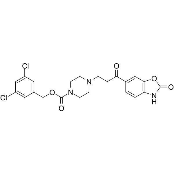
PF-8380 纯度: 98.95%
PF-8380 是一种有效的 autotaxin 抑制剂,体外酶实验和人类全血细胞实验中,IC50 分别为 2.8 nM 和 101 nM。

PF-8380 Chemical Structure
CAS No. : 1144035-53-9
| 规格 | 价格 | 是否有货 | 数量 |
|---|---|---|---|
| Free Sample (0.1-0.5 mg) | Apply now | ||
| 10 mM * 1 mL in DMSO | ¥704 | In-stock | |
| 5 mg | ¥640 | In-stock | |
| 10 mg | ¥900 | In-stock | |
| 50 mg | ¥3300 | In-stock | |
| 100 mg | ¥5900 | In-stock | |
| 200 mg | 询价 | ||
| 500 mg | 询价 |
* Please select Quantity before adding items.
PF-8380 相关产品
•相关化合物库:
- Bioactive Compound Library Plus
- Immunology/Inflammation Compound Library
- Metabolism/Protease Compound Library
- Anti-Cancer Compound Library
- Peptidomimetic Library
- Anti-Alzheimer’s Disease Compound Library
- Neurodegenerative Disease-related Compound Library
- Anti-Colorectal Cancer Compound Library
| 生物活性 |
PF-8380 is a potent autotaxin inhibitor with an IC50 of 2.8 nM in isolated enzyme assay and 101 nM in human whole blood. |
||||||||||||||||
|---|---|---|---|---|---|---|---|---|---|---|---|---|---|---|---|---|---|
| IC50 & Target[1] |
|
||||||||||||||||
| 体外研究 (In Vitro) |
PF-8380 also inhibits rat autotaxin with an IC50 of 1.16 nM with FS-3 substrate. Potency of PF-8380 is maintained when using enzyme produced from fetal fibroblasts used in combination with lysophosphatidyl choline (LPC) as a substrate. In human whole blood incubated with PF-8380 for 2 h, autotaxin is inhibited with an IC50 of 101 nM[1]. Autotaxin (ATX), an enzyme with lysophospholipase D (lysoPLD) activity, catalyzes the production of lysophosphatidic acid (LPA) from lysophosphatidylcholine (LPC). Pre-treatment of GL261 and U87-MG cells with 1 μM PF-8380 followed by 4 Gy irradiation results in decreased clonogenic survival, decreases migration (33% in GL261; P=0.002 and 17.9% in U87-MG; P=0.012), decreases invasion (35.6% in GL261; P=0.0037 and 31.8% in U87-MG; P=0.002), and attenuates radiation-induced Akt phosphorylation[2]. 上海金畔生物科技有限公司 has not independently confirmed the accuracy of these methods. They are for reference only. |
||||||||||||||||
| 体内研究 (In Vivo) |
The pharmacokinetic profile of PF-8380 is evaluated at an intravenous dose of 1 mg/kg and oral doses of 1 to 100 mg/kg out to 24 h. PF-8380 has mean clearance of 31 mL/min/kg, volume of distribution at steady state of 3.2 L/kg, and effective t1/2 of 1.2 h. Oral bioavailability is moderate, ranging from 43 to 83%. Plasma concentrations increased with single oral escalating doses, but Cmax increased at a rate that is approximately proportional to dose from 1 to 10 mg/kg and less than proportional to dose from 10 to 100 mg/kg. PF-8380 exposures estimated by area under the curve are approximately proportional to dose and linear up to 100 mg/kg. Plasma C16:0, C18:0, and C20:0 LPA levels are measured immediately after collection. Maximal reduction of LPA levels is observed by the 3 mg/kg dose at 0.5 h with all LPA returning at or above baseline at 24 h[1]. Treatment with 10 mg/kg PF-8380 increases tumor-associated vascularity modestly by 20% (P=0.497). When compared to control, treatment of PF-8380 45 min before 4 Gy irradiation decreases vascularity by nearly 48% when compared to control (P=0.031) and by 65% when compared to mice that received radiation alone (P=0.011)[2]. 上海金畔生物科技有限公司 has not independently confirmed the accuracy of these methods. They are for reference only. |
||||||||||||||||
| 分子量 |
478.33 |
||||||||||||||||
| Formula |
C22H21Cl2N3O5 |
||||||||||||||||
| CAS 号 |
1144035-53-9 |
||||||||||||||||
| 运输条件 |
Room temperature in continental US; may vary elsewhere. |
||||||||||||||||
| 储存方式 |
|
||||||||||||||||
| 溶解性数据 |
In Vitro:
DMSO : 6.67 mg/mL (13.94 mM; ultrasonic and adjust pH to 5 with HCl) 配制储备液
*
请根据产品在不同溶剂中的溶解度选择合适的溶剂配制储备液;一旦配成溶液,请分装保存,避免反复冻融造成的产品失效。 In Vivo:
请根据您的实验动物和给药方式选择适当的溶解方案。以下溶解方案都请先按照 In Vitro 方式配制澄清的储备液,再依次添加助溶剂: ——为保证实验结果的可靠性,澄清的储备液可以根据储存条件,适当保存;体内实验的工作液,建议您现用现配,当天使用; 以下溶剂前显示的百
|
||||||||||||||||
| 参考文献 |
|
| Kinase Assay [1] |
FS-3 substrate is solubilized in assay buffer at 500 μM and frozen at -20°C in single-use aliquots for up to 4 weeks. Recombinant autotaxin is diluted in Tris-buffered saline (140 mM NaCl, 5 mM KCl, 1 mM CaCl2, 1 mM MgCl2, 50 mM Tris, pH 8.0) and incubated with compound in DMSO or DMSO alone (final 1% DMSO) for 15 min at 37°C, and the reaction is started with the addition of FS-3 at a final concentration of 1 μM. The reaction is allowed to proceed at 37°C for 30 min and monitored at 520 nm until the uninhibited control compared with a no-enzyme control gave a Z′≥0.5. IC50s are determined in triplicate by using a four-parameter fit[1]. 上海金畔生物科技有限公司 has not independently confirmed the accuracy of these methods. They are for reference only. |
|---|---|
| Cell Assay [2] |
HUVEC (1×10⁶) and bEnd.3 cells (1×10⁶) are plated in 100 mm plates and after 24 h, U87-MG (2×10⁶) and GL261 (2×10⁶) cells are plated onto transwell inserts. After co-culture for 24 h, cells are treated with 1 μM of PF-8380 or vehicle control DMSO for 45 min prior to irradiation with 0, 2, 4, 6, or 8 Gy. After the treatments as co-culture with either PF-8380 or DMSO calculated numbers of U87-MG and GL261 cells are plated to enable normalization for plating efficiencies. After 7 to 10 day incubation plates are fixed with 70% EtOH and stained with 1% methylene blue. Colonies consisting of >50 cells are counted by viewing the plates under a microscope. The survival fractions are calculated as (number of colonies/number of cells plated)/(number of colonies for corresponding control/number of cells plated). Survival curves are analyzed by curve fitting to the alpha/beta model calculating D0 and n[2]. 上海金畔生物科技有限公司 has not independently confirmed the accuracy of these methods. They are for reference only. |
| Animal Administration [1][2] |
Rats[1] 上海金畔生物科技有限公司 has not independently confirmed the accuracy of these methods. They are for reference only. |
| 参考文献 |
|
所有产品仅用作科学研究或药证申报,我们不为任何个人用途提供产品和服务
