上海金畔生物科技有限公司为生命科学和医药研发人员提供生物活性分子抑制剂、激动剂、特异性抑制剂、化合物库、重组蛋白,专注于信号通路和疾病研究领域。
PRT062607 Hydrochloride (Synonyms: P505-15 Hydrochloride) 纯度: 98.06%
PRT062607 Hydrochloride (P505-15 Hydrochloride) 是一种有效的纯化的 Syk 抑制剂,IC50 为 1-2 nM。

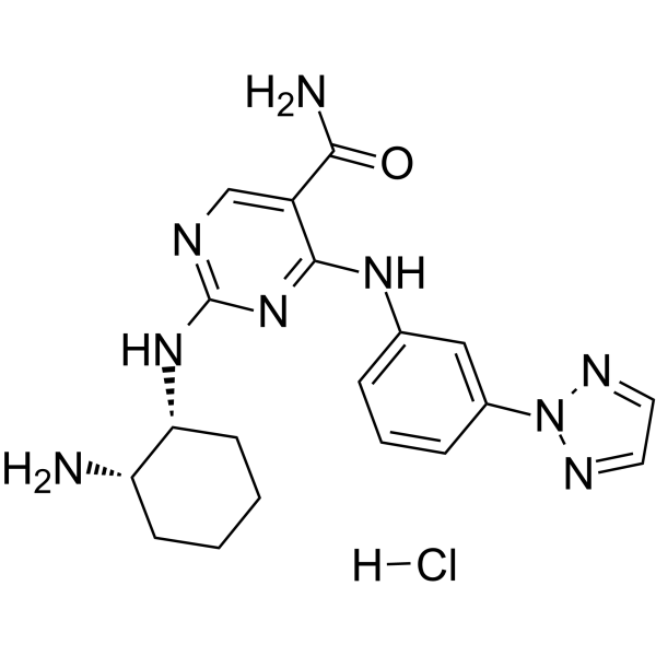
PRT062607 Hydrochloride Chemical Structure
CAS No. : 1370261-97-4
| 规格 | 价格 | 是否有货 | 数量 |
|---|---|---|---|
| Free Sample (0.1-0.5 mg) | Apply now | ||
| 10 mM * 1 mL in DMSO | ¥1377 | In-stock | |
| 5 mg | ¥1252 | In-stock | |
| 10 mg | ¥1841 | In-stock | |
| 50 mg | ¥6535 | In-stock | |
| 100 mg | 询价 | ||
| 200 mg | 询价 |
* Please select Quantity before adding items.
PRT062607 Hydrochloride 相关产品
•相关化合物库:
- Drug Repurposing Compound Library Plus
- Clinical Compound Library Plus
- Bioactive Compound Library Plus
- Kinase Inhibitor Library
- Protein Tyrosine Kinase Compound Library
- Anti-Cancer Compound Library
- Clinical Compound Library
- Drug Repurposing Compound Library
- Chemical Probe Library
| 生物活性 |
PRT062607 Hydrochloride (P505-15 Hydrochloride) is a highly specific and potent inhibitor of purified Syk (IC50 1-2 nM). |
IC50 & Target |
IC50: 1 nM (Syk), 81 nM (Fgr), 88 nM (MLK1), 123 nM (Yes)[1] |
||||||||||||||
|---|---|---|---|---|---|---|---|---|---|---|---|---|---|---|---|---|---|
| 体外研究 (In Vitro) |
PRT062607 (P505-15) is a novel, highly specific, and potent orally available small-molecule inhibitor of Syk. The potency of PRT062607 against its target kinase Syk is initially tested in two different purified kinase assays. Using a FRET assay, half-maximal Syk inhibition required 6±0.2 nM (mean±S.E.M.). Similar potency is observed when tested in a radioactive enzyme assay, with a resulting Syk IC50 of 2.1±0.4 nM (mean±S.E.M.). In human whole blood, PRT062607 potently inhibits B cell antigen receptor-mediated B cell signaling and activation (IC50 0.27 and 0.28 μM, respectively) and Fcε receptor 1-mediated basophil degranulation (IC50 0.15 μM)[1]. 上海金畔生物科技有限公司 has not independently confirmed the accuracy of these methods. They are for reference only. |
||||||||||||||||
| 体内研究 (In Vivo) |
In the mouse CAIA model, oral administration of PRT062607 (P505-15) results in an average inhibition of paw inflammation, as measured by daily scoring of inflammation compared with vehicle controls, of 12, 44, and 87% with average plasma concentration (C average over 24 h) assessed at the end of the study of 0.38, 0.95, and 1.47 μM, respectively. In mice treated with 30 mg/kg PRT062607, the damage to the joints is significantly reduced and seemed indistinguishable from normal mice. In the rat CIA model, the high dose of PRT062607 (15 mg/kg b.i.d.) completely suppresses inflammation in a majority of the animals (seven of eight), by the end of the study (mean inflammation score±S.E.M.=0.63±1.1; p<0.0001 versus vehicle)[1]. 上海金畔生物科技有限公司 has not independently confirmed the accuracy of these methods. They are for reference only. |
||||||||||||||||
| Clinical Trial |
|
||||||||||||||||
| 分子量 |
429.91 |
||||||||||||||||
| Formula |
C19H24ClN9O |
||||||||||||||||
| CAS 号 |
1370261-97-4 |
||||||||||||||||
| 运输条件 |
Room temperature in continental US; may vary elsewhere. |
||||||||||||||||
| 储存方式 |
4°C, sealed storage, away from moisture *In solvent : -80°C, 6 months; -20°C, 1 month (sealed storage, away from moisture) |
||||||||||||||||
| 溶解性数据 |
In Vitro:
H2O : ≥ 50 mg/mL (116.30 mM) DMSO : ≥ 33 mg/mL (76.76 mM) * “≥” means soluble, but saturation unknown. 配制储备液
*
请根据产品在不同溶剂中的溶解度选择合适的溶剂配制储备液;一旦配成溶液,请分装保存,避免反复冻融造成的产品失效。 In Vivo:
请根据您的实验动物和给药方式选择适当的溶解方案。以下溶解方案都请先按照 In Vitro 方式配制澄清的储备液,再依次添加助溶剂: ——为保证实验结果的可靠性,澄清的储备液可以根据储存条件,适当保存;体内实验的工作液,建议您现用现配,当天使用; 以下溶剂前显示的百
|
||||||||||||||||
| 参考文献 |
|
| Kinase Assay [1] |
Potency of Syk inhibition is determined by using a fluorescence resonance energy transfer (FRET) assay. The extent of substrate phosphorylation by Syk is measured in the presence of various PRT062607 (P505-15) concentrations. Syk activity is determined by a fluorescent antibody specific for phosphorylated tyrosine by using the increase of FRET. Twelve concentrations are tested for dose response. Specificity and potency of kinase inhibition is determined by evaluation of PRT062607 in the Kinase Profiler panel of 270 independent purified kinase assays. For profiling, PRT062607 is tested in duplicate at two concentrations at a fixed concentration of ATP. Subsequently, IC50 determinations using the radioactive assays are carried out at an ATP concentration optimized for each individual kinase. All radioactive ATP incorporation enzyme assays are performed[1]. 上海金畔生物科技有限公司 has not independently confirmed the accuracy of these methods. They are for reference only. |
|---|---|
| Cell Assay [1] |
SUDHL4 cells (106 cells) are stimulated for 30 min with F(ab)′2 anti-human IgM antibody. Signaling is terminated by resuspending cells in ice-cold lysis buffer (50 mM Tris, pH 7.4, 150 mM NaCl, 0.5 mM EDTA, 1% nonidet P-40, and 0.5% sodium deoxycholate) containing freshly added protease and phosphatase inhibitor tablets. Protein lysates are resolved by SDS-polyacrylamide gel electrophoresis under reducing conditions and probed with the indicated antibodies. Secondary antibody conjugated to horseradish peroxidase and enhanced chemiluminescence detection reagent are used for the detection of specific protein. Where indicated, cells are pretreated for 1 h at 37°C with PRT062607 or vehicle control before stimulation. Densitometry of exposed films is performed by using the AlphaImager 2200. Ba/F3 cells are seeded at 5000 cells per well in a 384-well tissue culture plate containing media with various concentrations of PRT062607 or staurosporin. After 3 days, viability of cells is quantitated by CellTiter Glo following the manufacturer-supplied protocols[1]. 上海金畔生物科技有限公司 has not independently confirmed the accuracy of these methods. They are for reference only. |
| Animal Administration [1] |
Mice[1] 上海金畔生物科技有限公司 has not independently confirmed the accuracy of these methods. They are for reference only. |
| 参考文献 |
|
所有产品仅用作科学研究或药证申报,我们不为任何个人用途提供产品和服务
