上海金畔生物科技有限公司为生命科学和医药研发人员提供生物活性分子抑制剂、激动剂、特异性抑制剂、化合物库、重组蛋白,专注于信号通路和疾病研究领域。
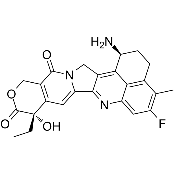
Exatecan (Synonyms: 依喜替康; DX-8951)
Exatecan (DX-8951) 是一种 DNA 拓扑异构酶 I (topoisomerase I) 抑制剂,IC50 值为 2.2 μM (0.975 μg/mL),可用于癌症研究。

Exatecan Chemical Structure
CAS No. : 171335-80-1
| 规格 | 是否有货 | ||
|---|---|---|---|
| 100 mg | 询价 | ||
| 250 mg | 询价 | ||
| 500 mg | 询价 |
* Please select Quantity before adding items.
Exatecan 的其他形式现货产品:
| 生物活性 |
Exatecan (DX-8951) is a DNA topoisomerase I inhibitor, with an IC50 of 2.2 μM (0.975 μg/mL), and can be used in cancer research. |
|
|---|---|---|
| IC50 & Target[1] |
|
|
| 体外研究 (In Vitro) |
Exatecan is a potent topoisomerase I inhibitor, with an IC50 of 0.975 μg/mL. Exatecan Mesylate (DX-8951f) significantly inhibits the proliferation of several cancer cell lines, with mean GI50s of 2.02 ng/mL, 2.92 ng/mL, 1.53 ng/mL, and 0.877 ng/mL for breast cancer cells, colon cancer cells, stomach cancer cells and lung cancer cells, respectively[1]. Exatecan Mesylate (DX-8951f) displays cytotoxic activities against PC-6, PC-6/SN2-5 cells, with mean GI50s of 0.186 and 0.395 ng/mL, respctively. Exatecan Mesylate (34 nM) stabilizes DNA-TopoI complexes in PC-6 and PC-6/SN2-5 cells[3]. Shanghai Jinpan Biotech Co Ltd has not independently confirmed the accuracy of these methods. They are for reference only. |
|
| 体内研究 (In Vivo) |
Exatecan Mesylate (DX-8951f, 3.325-50 mg/kg, i.v.) exhibits antitumor activities in the mice model bearing tumor cells, without toxic death[1]. Exatecan Mesylate (15, 25 mg/kg, i.v.) hightly inhibits MIA-PaCa, BxPC-3 primary tumor growth in the MIA-PaCa-2 early-stage model and early-stage model of BxPC-3. Exatecan Mesylate (15, 25 mg/kg, i.v.) also significantly suppresses BxPC-3 lymphatic metastasis and completely eliminates lung metastasis in the BxPC-3 late-stage cancer model[2]. Shanghai Jinpan Biotech Co Ltd has not independently confirmed the accuracy of these methods. They are for reference only. |
|
| Clinical Trial |
|
|
| 分子量 |
435.45 |
|
| Formula |
C24H22FN3O4 |
|
| CAS 号 |
171335-80-1 |
|
| 中文名称 |
依喜替康;依沙替康 |
|
| 运输条件 |
Room temperature in continental US; may vary elsewhere. |
|
| 储存方式 |
-20°C, sealed storage, away from moisture *In solvent : -80°C, 6 months; -20°C, 1 month (sealed storage, away from moisture) |
|
| 参考文献 |
|
| Kinase Assay |
sup>[3]Cells (5×10⁶) are lysed with SDS buffer (10 mM HEPES, 2 mM orthovanadate, 10 mM NaF, 10 mM pyrophosphate, 1 mMPMSF, 10 µg/mL leupeptin, 10% 2-mercaptoethanol, 10% glycerol,8% SDS, 42 mM Tris-HCl, 0.002% bromophenol blue, pH 7.4). Protein in the whole cell lysates is separated in 7.5% polyacryl-amide gel and blotted onto nitrocellulose membrane. The membrane is treated with anti-Topo I human antibody and subsequently, with horseradish peroxidase-conjugated protein A. The Topo I-specific band is detected with ECL reagents. To obtain a nuclear extract, cells (5×10⁷) are washed with ice-cold buffer (2 mM K2HPO4, 5 mM MgCl2, 150 mM NaCl, 1 mM EGTA, 0.1 mM dithiothreitol), resuspended in buffer containing 0.35% Triton-X100 and PMSF and then incubated on ice for 10 min. The resulting lysates are centrifuged, and precipitates are then incubated with buffer containing 0.35 M NaCl for 1 hr at 4°C. After centrifugation (18,000g, 10 min), the protein concentration of the supernatant (nuclear extract) is determined by Bradford’s method using a protein assay kit. The same amount of nuclear protein is analyzed by Western blotting analysis using anti-Topo I antibody[3]. Shanghai Jinpan Biotech Co Ltd has not independently confirmed the accuracy of these methods. They are for reference only. |
|---|---|
| Cell Assay [1] |
Growth inhibition experiments are carride out in 96-well flat-bottomed microplates, and the amount of viable cell at the end of the incubation is determined by MTT assay. Thus, 500-20000 cells/well in 150 μL of medium are plated and grown for 24 h (P388, CCRF-CEM and K562 cells for 4h), the drug (including Exatecan Mesylate, in 150 μL medium/well), or the medium alone as a control, is added, and the cells are cultured for an additional 3 days. After addition of MTT (20 μL/well, 5 mg/mL in phosphate-buffered saline), the plates are incubated for 4 h and centrifuged at 800 g for 5 min, then the medium is removed and the blue dye formed is dissolved in 150 μL of DMSO. the absorbance is measured at 540 nm using a Microplate Reader model 3550[1]. Shanghai Jinpan Biotech Co Ltd has not independently confirmed the accuracy of these methods. They are for reference only. |
| Animal Administration [2] |
At 3 weeks after BxPC-3-GFP and MIA-PaCa-2-GFP orthotopic implantation, mice are randomized into five different groups of 5 mice each for treatment purposes. Group 1 serves as the negative control and does not receive any treatment. Groups 2 and 3 are treated with Exatecan Mesylate at 25 and 15 mg/kg/dose, respectively. Groups 4 and 5 receive gemcitabine treatments at 300 and 150 mg/kg/dose, respectively. At 6 weeks after BxPC-3-GFP orthotopic implantation, mice are randomized into three different groups of 20 mice each for treatment purposes. Group 1 serves as the negative control and does not receive any treatment. Group 2 is treated with 25 mg/kg/dose Exatecan Mesylate and group 3 receives 300 mg/kg/dose gemcitabine. Dosing for both drugs is performed once a week for 3 weeks, discontinued for 2 weeks, and then continued for another 3 weeks. In both early and late cancer models, primary tumor size and body weights are measured once a week. Tumor volumes are calculated using the formula a × b2 × 0.5, where a and b represent the larger and smaller diameters, respectively. At the termination of the studies, mice are sacrificed and explored. Final tumor weights and direct GFP images of primary tumor and metastases are recorded for each mouse. The tumor growth IR is calculated using the formula IR (%) = (1 − TWt/TWc) × 100, where TWt and TWc are the mean tumor weight of treated and control groups, respectively[2]. Shanghai Jinpan Biotech Co Ltd has not independently confirmed the accuracy of these methods. They are for reference only. |
| 参考文献 |
|
所有产品仅用作科学研究或药证申报,我们不为任何个人用途提供产品和服务
