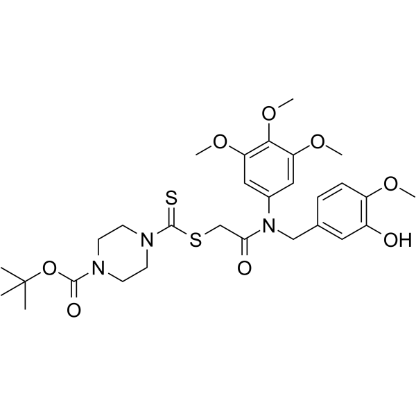
体外研究
(In Vitro) |
Tubulin polymerization-IN-5 (compound 20q) (0-100 nM, 48 h) exhibits potent antiproliferative activity against Kyse30, Kyse450, MGC-803, and HCT-116 cells[1].
Tubulin polymerization-IN-5 (0-60 nM, 7 d) obviously inhibits the cellular colony formation ability of the Kyse30 and Kyse450 cells[1].
Tubulin polymerization-IN-5 (0-100 nM, 48 h) inhibits microtubule assembly and disrupts cytoskeleton[1].
Tubulin polymerization-IN-5 (0-300 nM, 24 h) causes a significant weakening of the β-tubulin adduct band in Kyse30 and Kyse450 cells, competitively bind the colchicine binding site of β-tubulin[1].
Tubulin polymerization-IN-5 (0-100 nM, 48 h) effectively arrests cells at the G2/M phase, and induces cell apoptosis in Kyse30 and Kyse450 cells by regulating the expression of related proteins[1].
Tubulin polymerization-IN-5 (0-100 nM, 48 h) induces cell mitochondrial apoptosis in ESCC cells, leads to a significant depolarization of mitochondria membrane potential in Kyse30 and Kyse450 cells[1].
Shanghai Jinpan Biotech Co Ltd has not independently confirmed the accuracy of these methods. They are for reference only.
Cell Proliferation Assay
| Cell Line: |
Kyse30, Kyse450, MGC-803, and HCT-116 cells[1] |
| Concentration: |
0, 80, 100 nM |
| Incubation Time: |
48 h |
| Result: |
Exhibited potent antiproliferative activity against Kyse30, Kyse450, MGC-803, and HCT-116 cells, with IC50 values of 0.069, 0.078, 0.084, and 0.227 μM, respectively. |
Cell Cycle Analysis
| Cell Line: |
Kyse30 and Kyse450 cells[1] |
| Concentration: |
0, 80, 100 nM |
| Incubation Time: |
48 h |
| Result: |
Effectively arrested 2 ESCC cells at the G2/M phase, significantly increased of percentages of cells at the G2/M phase from 19.38 to 76.9767 in Kyse30 cells, and 7.04333 to 80.8933 in Kyse450 cells. |
Apoptosis Analysis
| Cell Line: |
Kyse30 and Kyse450 cells[1] |
| Concentration: |
0, 80, 100 nM |
| Incubation Time: |
48 h |
| Result: |
Dose-dependently induced cell apoptosis in Kyse30 and Kyse450 cells, significantly increased the percentages of total apoptotic cells from 8.1667% (0 nM) to 23.8% (80 nM, Kyse30 cells), 34.0333% (80 nM, Kyse450 cells), 38.633% (100 nM, Kyse30 cells), and 57.3667% (100 nM, Kyse450 cells), respectively. |
Western Blot Analysis
| Cell Line: |
Kyse30 and Kyse450 cells[1] |
| Concentration: |
0, 80, 100 nM |
| Incubation Time: |
48 h |
| Result: |
Dose-dependently down-regulated the expression of the G2 phase related proteins CDK1, CDC25c and p-Wee 1, up-regulated the level of the M phase marker protein p-Histone H3; up-regulated the activity of the executors of apoptosis caspase-3, up-regulated the pro-apoptotic proteins Bax and Noxa, and down-regulated the anti-apoptotic protein Bcl-2, decreased the levels of IAP (Inhibitor of Apoptosis Proteins) family protein XIAP. |
|

