上海金畔生物科技有限公司为生命科学和医药研发人员提供生物活性分子抑制剂、激动剂、特异性抑制剂、化合物库、重组蛋白,专注于信号通路和疾病研究领域。
IOX2 纯度: 99.71%
IOX2 是一种特异性的脯氨酰羟化酶-2 (PHD2) 抑制剂,IC50 为 22 nM。

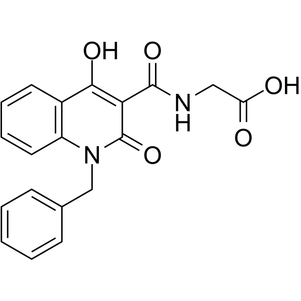
IOX2 Chemical Structure
CAS No. : 931398-72-0
| 规格 | 价格 | 是否有货 | 数量 |
|---|---|---|---|
| Free Sample (0.1-0.5 mg) | Apply now | ||
| 10 mM * 1 mL in DMSO | ¥778 | In-stock | |
| 5 mg | ¥707 | In-stock | |
| 10 mg | ¥1083 | In-stock | |
| 50 mg | ¥4201 | In-stock | |
| 100 mg | 询价 | ||
| 200 mg | 询价 |
* Please select Quantity before adding items.
IOX2 相关产品
•相关化合物库:
- Bioactive Compound Library Plus
- Epigenetics Compound Library
- Immunology/Inflammation Compound Library
- Metabolism/Protease Compound Library
- Anti-Cancer Compound Library
- Reprogramming Compound Library
- Oxygen Sensing Compound Library
- Chemical Probe Library
- Glutamine Metabolism Compound Library
- Anti-Breast Cancer Compound Library
- Anti-Cancer Metabolism Compound Library
- Angiogenesis Related Compound Library
- Transcription Factor Targeted Library
| 生物活性 |
IOX2 is a specific prolyl hydroxylase-2 (PHD2) inhibitor with IC50 of 22 nM. |
IC50 & Target |
IC50: 22 nM (PHD2)[1] |
||||||||||||||
|---|---|---|---|---|---|---|---|---|---|---|---|---|---|---|---|---|---|
| 体外研究 (In Vitro) |
IOX2 is at least 2-5000 fold selective, as judged by IC50 values, for PHD2 over the KDMs and Factor Inhibiting HIF(FIH)[1]. IOX2 significantly upregulates the transcription of VEGF-A and BNIP3 in normal human epidermal keratinocytes (NHEK) and normal human dermal fibroblasts (NHDF) when grown under normoxia and hypoxia. IOX2 efficiently promotes HIF-1α stability, nuclear translocation, and target gene expression in keratinocytes and fibroblasts. In addition, IOX2 significantly upregulates biosynthesis and transcription of VEGF-A and BNIP3 in sulfur mustard (SM)-exposed NHEK and NHDF grown under hypoxia. These results suggest that application of IOX2 is useful for restoring of SM-affected HIF-1α stability and signaling activity in keratinocytes and fibroblasts[2]. 上海金畔生物科技有限公司 has not independently confirmed the accuracy of these methods. They are for reference only. |
||||||||||||||||
| 体内研究 (In Vivo) |
To investigate the utility of IOX2 as in vivo functional probes, IOX2 is tested to upregulate HIF signaling in a whole organism, that is, transgenic zebrafish (Danio rerio). Because the expression of the PHD3 encoding gene is regulated by HIF in humans and zebrafish, PHD3 levels are a readout of HIF activity. A zebrafish hypoxia reporter line is generated expressing GFP with the phd3 promoter elements. Transgenic wild-type embryos at 3 days postfertilization treated with compounds (10 μM) for 2 days displayed clear increase in phd3:EGFP expression in the liver, relative to controls. Significant increases in GFP levels are observed with IOX2[1]. 上海金畔生物科技有限公司 has not independently confirmed the accuracy of these methods. They are for reference only. |
||||||||||||||||
| 分子量 |
352.34 |
||||||||||||||||
| Formula |
C19H16N2O5 |
||||||||||||||||
| CAS 号 |
931398-72-0 |
||||||||||||||||
| 运输条件 |
Room temperature in continental US; may vary elsewhere. |
||||||||||||||||
| 储存方式 |
|
||||||||||||||||
| 溶解性数据 |
In Vitro:
DMSO : 25 mg/mL (70.95 mM; Need ultrasonic) 配制储备液
*
请根据产品在不同溶剂中的溶解度选择合适的溶剂配制储备液;一旦配成溶液,请分装保存,避免反复冻融造成的产品失效。 In Vivo:
请根据您的实验动物和给药方式选择适当的溶解方案。以下溶解方案都请先按照 In Vitro 方式配制澄清的储备液,再依次添加助溶剂: ——为保证实验结果的可靠性,澄清的储备液可以根据储存条件,适当保存;体内实验的工作液,建议您现用现配,当天使用; 以下溶剂前显示的百
|
||||||||||||||||
| 参考文献 |
|
| Kinase Assay [1] |
Inhibition assays are carried out in 384-well white ProxiPlates in 10 μL of reaction volume. Standard reaction mixtures consisted of the compound (in 2% DMSO final concentration), enzyme mix (0.001 μM of PHD2, 10 μM of Fe(II), 100 μM of ascorbate) and peptide mix (0.06 μM of biotinylated C-terminal oxygen dependent degradation domain (CODD) peptide, 2 μM of 2OG) in 50 mM HEPES pH 7.5, 0.01% Tween-20 and 0.1% BSA buffer. Compounds (e.g., IOX2) are preincubated with the enzyme mix for 15 min before being incubated with peptide mix for 10 min at 22°C. Each reaction is quenched with 5 μL of 30 mM EDTA. The bead mix containing AlphaScreen beads is preincubated for 1h with a rabbit monoclonal antibody selective for hydroxy-HIF1α (Pro564) and are added to the wells for a further 1 h at 22°C. The plates are then analyzed with an Envision plate reader. The IC50 values are calculated using nonlinear regression with normalized dose-response fit using Prism GraphPad (n≥3)[1]. 上海金畔生物科技有限公司 has not independently confirmed the accuracy of these methods. They are for reference only. |
|---|---|
| Cell Assay [1] |
Both VHL-defective (renal carcinoma cells with an empty vector, RCC4) and VHL-competent cells human embryonic kidney HEK293T, osteosarcoma U2OS and RCC4/VHLHA (RCC4 stably transfected with C-terminal HA-tagged wt VHL) are used. Cells are treated with DMSO (control) and tested compounds (e.g., IOX2) (dissolved in DMSO except for DMOG which is dissolved in PBS and added directly to culture medium) for 4-5 h. Cell extracts are probed with antibodies to hydroxy-Pro564 (CODD-OH) and hydroxy-Asn803 (CAD-OH). HIF-1α band intensities are used to normalize hydroxylation signals[1]. 上海金畔生物科技有限公司 has not independently confirmed the accuracy of these methods. They are for reference only. |
| Animal Administration [1] |
Zebrafish[1] 上海金畔生物科技有限公司 has not independently confirmed the accuracy of these methods. They are for reference only. |
| 参考文献 |
|
所有产品仅用作科学研究或药证申报,我们不为任何个人用途提供产品和服务
