上海金畔生物科技有限公司为生命科学和医药研发人员提供生物活性分子抑制剂、激动剂、特异性抑制剂、化合物库、重组蛋白,专注于信号通路和疾病研究领域。
KN-62 纯度: 99.45%
KN-62 是一种选择性的、可逆的钙调蛋白依赖性蛋白激酶 II (CaMK-II) 抑制剂,对大鼠脑 CaMK-II 的 IC50 值为 0.9 μM。KN-62 直接与 CaMK-II 酶的钙调蛋白结合位点结合。KN-62 是非竞争性的 P2X7 受体拮抗剂,IC50 约为 15 nM。

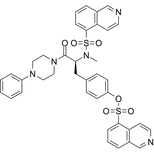
KN-62 Chemical Structure
CAS No. : 127191-97-3
| 规格 | 价格 | 是否有货 | 数量 |
|---|---|---|---|
| 10 mM * 1 mL in DMSO | ¥1270 | In-stock | |
| 5 mg | ¥800 | In-stock | |
| 10 mg | ¥1360 | In-stock | |
| 50 mg | ¥5500 | In-stock | |
| 100 mg | 询价 | ||
| 200 mg | 询价 |
* Please select Quantity before adding items.
KN-62 相关产品
•相关化合物库:
- Covalent Screening Library Plus
- Bioactive Compound Library Plus
- Kinase Inhibitor Library
- Membrane Transporter/Ion Channel Compound Library
- Neuronal Signaling Compound Library
- Anti-Cancer Compound Library
- Covalent Screening Library
- Neurotransmitter Receptor Compound Library
| 生物活性 |
KN-62 is a selective and reversible inhibitor of calmodulin-dependent protein kinase II (CaMK-II) with a Ki of 0.9 μM for rat brain CaMK-II. KN-62 directly binds to the calmodulin binding site of CaMK-II. KN-62 displays noncompetitive antagonism at P2X7 receptors in HEK293 cells, with an IC50 value of approximately 15 nM. |
||||||||||||||||
|---|---|---|---|---|---|---|---|---|---|---|---|---|---|---|---|---|---|
| IC50 & Target[1][2] |
|
||||||||||||||||
| 体外研究 (In Vitro) |
KN-62 potently antagonizes ATP-stimulated Ba2+ influx into fura-2 loaded human lymphocytes with an IC50 of 12.7 nM and complete inhibition of the flux at a concentration of 500 nM[1]. 上海金畔生物科技有限公司 has not independently confirmed the accuracy of these methods. They are for reference only. |
||||||||||||||||
| 体内研究 (In Vivo) |
KN62 (5 mg/kg/day; ip; three times a week for 6 weeks) significantly reduces the liver metastatic tumor burden in five weeks old BALB/c athymic nude mice inoculated with TAMR-MCF-7 cells[3]. 上海金畔生物科技有限公司 has not independently confirmed the accuracy of these methods. They are for reference only. |
||||||||||||||||
| 分子量 |
721.84 |
||||||||||||||||
| Formula |
C38H35N5O6S2 |
||||||||||||||||
| CAS 号 |
127191-97-3 |
||||||||||||||||
| 运输条件 |
Room temperature in continental US; may vary elsewhere. |
||||||||||||||||
| 储存方式 |
|
||||||||||||||||
| 溶解性数据 |
In Vitro:
DMSO : ≥ 100 mg/mL (138.53 mM) * “≥” means soluble, but saturation unknown. 配制储备液
*
请根据产品在不同溶剂中的溶解度选择合适的溶剂配制储备液;一旦配成溶液,请分装保存,避免反复冻融造成的产品失效。 In Vivo:
请根据您的实验动物和给药方式选择适当的溶解方案。以下溶解方案都请先按照 In Vitro 方式配制澄清的储备液,再依次添加助溶剂: ——为保证实验结果的可靠性,澄清的储备液可以根据储存条件,适当保存;体内实验的工作液,建议您现用现配,当天使用; 以下溶剂前显示的百
|
||||||||||||||||
| 参考文献 |
|
| Kinase Assay [1] |
Lymphocytes (1×10⁷/mL) are cultured with [3H]-oleic acid (2-5 μCi/mL, specific activity 10 Ci/mmol) for 20-24 h in RPMI-1640 medium supplemented with Gentamicin (40 μg/mL), 10% heat inactivated foetal calf serum (FCS) at 37°C to label membrane phospholipids. Labelled cells are washed twice in HEPES buffered saline followed by a final wash in either HEPES buffered saline or 150 mM KCl medium containing HEPES 10 mM, pH 7.4, bovine serum albumin (BSA) 1 g/L and D-glucose 5 mM and CaCl2 1 mM. Three mL aliquots containing 1.1×10< sup>7/mL lymphocytes are warmed to 37°C and incubated with or without KN-62 or KN-04 (1 nM-500 nM) for 5 min, then 900 mL aliquots are added to 100 uL butanol (final concentration 30 mM) for a further 5 min, and stimulated with 1 mM ATP for 15 min with gentle mixing in the continued presence of inhibitor or diluent. The phospholipase D reaction is terminated by addition of 1 mL of 20 mM MgCl2 followed by centrifugation and addition of 1 mL ice cold methanol. Membrane lipids are extracted into chloroform/HCl at 4°C under N2, and separated by silica gel thin layer chromatography (t.l.c.) with the solvent system, ethyl acetate/iso-octane/acetic acid/water (13:2:3:10, v/v) under saturating conditions. Sample spots are located by autoradiography and [3H]-phosphatidylbutanol ([3H]-PBut) spots identified by an authentic standard. [3H]-PBut and [3H]-phospholipid spots are scraped into scintillant fluid (PPO in toluene, 4 g/L) and counted in a liquid scintillation counter. The quantity of [3H]-PBut is presented as a percentage of total 3H labelled-cellular phospholipids. Phospholipase D assays are performed in triplicate[1]. 上海金畔生物科技有限公司 has not independently confirmed the accuracy of these methods. They are for reference only. |
|---|---|
| Cell Assay [4] |
All experiments are performed using adherent HEK293 cells stably transfected with cDNA encoding the human P2X7 receptor. Adherent cells on 12-well polylysine-coated plates are incubated at 37°C in 1 mL physiological salt solution (125 mM NaCl, 5 mM KCl, 1 mM MgCl2, 1.5 mM CaCl2, 25 mM NaHEPES (pH 7.5), 10 mM D-glucose, 1 mg/mL BSA). Antagonists(e.g., KN-62) are added from 1,000× stock solutions dissolved in DMSO. Cells are preincubated with antagonists (e.g., KN-62) for 15 min prior to stimulation for 10 min with 3 mM ATP (final concentration). Reactions are terminated by rapid aspiration of the extracellular medium in each well. The adherent cells in each well are then extracted overnight with 1 mL 10% HNO3. K+ content in these nitric acid extracts is assayed by atomic absorbance spectrophotometry. Duplicate or triplicate wells are run for all test conditions in each separate experiment[4]. 上海金畔生物科技有限公司 has not independently confirmed the accuracy of these methods. They are for reference only. |
| Animal Administration [5] |
Mice[3] 上海金畔生物科技有限公司 has not independently confirmed the accuracy of these methods. They are for reference only. |
| 参考文献 |
|
所有产品仅用作科学研究或药证申报,我们不为任何个人用途提供产品和服务
