体外研究
(In Vitro) |
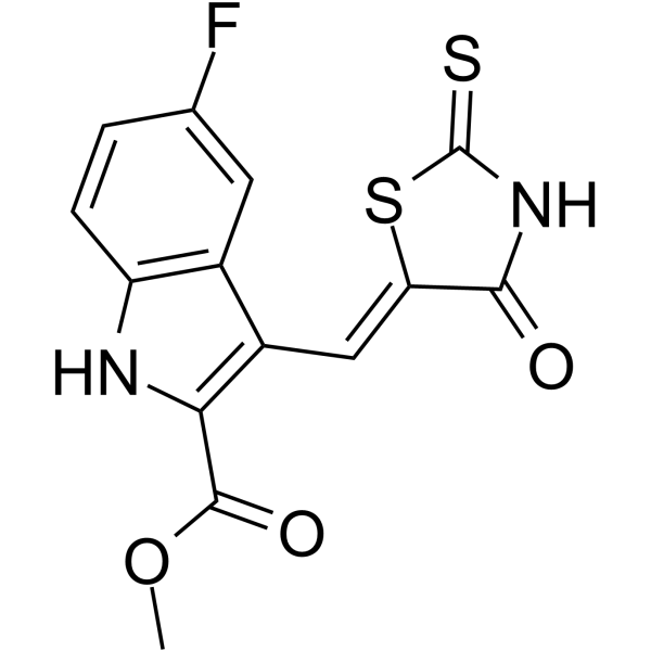
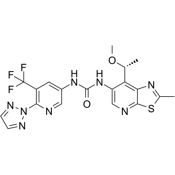
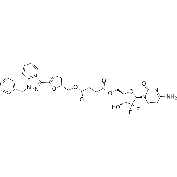
Anticancer agent 43 (compound 3a) shows selectivity toward human tumor cells (SI50=28.94)[1].
Anticancer agent 43 (45 µM, 24 h) induces apoptosis via caspase 3, PARP1 and Bax dependent pathways in HepG2 cells[1].
Anticancer agent 43 (45 µM, 24 h) shows no effect on the transition of G1/S phases in HepG2 cells[1].
Anticancer agent 43 (0.7, 45, 55 µM) induces DNA damage in HCT116 cells (Tail DNA=16.1%, OTM=3.7), MCF-7 cells, HepG2 cells (Tail DNA=26.2%, OTM=13.2), Balb/c 3T3 cells (Tail DNA = 8.4%, OTM = 3.5)[1].
Shanghai Jinpan Biotech Co Ltd has not independently confirmed the accuracy of these methods. They are for reference only.
Cell Cytotoxicity Assay[1]
| Cell Line: |
HepG2, MCF-7, HCT116, HeLa, A549, WM793, THP-1, HaCaT, Balb/c3T3 cells |
| Concentration: |
0, 1, 10, 100 µM |
| Incubation Time: |
72 h |
| Result: |
Showed cytotoxic action with GI50s of 12.1, 0.7, 0.8, 49.3, 9.7 µM for for HepG2, MCF-7, HCT116, HeLa, A549 cells, low toxicity towards WM793, THP-1, HaCaT, Balb/c 3T3 cells with GI50s of 80.4, 62.4,98.3,40.8 µM , respectively. |
Apoptosis Analysis[1]
| Cell Line: |
HepG2 cells |
| Concentration: |
45 µM |
| Incubation Time: |
24 h |
| Result: |
Induced apoptosis in HepG2 cells via caspase 3, PARP1 and Bax dependent pathways. |
Western Blot Analysis[1]
| Cell Line: |
HCT116, MCF-7 cells |
| Concentration: |
0.7 µM |
| Incubation Time: |
24 h |
| Result: |
Decreased the expression of Cdk2 protein in HCT116 and MCF-7 cells. |
Cell Cycle Analysis[1]
| Cell Line: |
HepG2 cells |
| Concentration: |
45 µM |
| Incubation Time: |
24 h |
| Result: |
Showed no effect on the transition of G1/S phases in HepG2 cells. |
|

![Splenopentin 编码 [75957-60-7]](http://www.fluoroprobe.com/wp-content/uploads/2022/10/20221006_633ed4f71ed31.png)
![Splenopentin 编码 [75957-60-7]](http://www.fluoroprobe.com/wp-content/uploads/2022/10/20221006_633ed4f778364.jpg)
