上海金畔生物科技有限公司为生命科学和医药研发人员提供生物活性分子抑制剂、激动剂、特异性抑制剂、化合物库、重组蛋白,专注于信号通路和疾病研究领域。
2-Methoxyestradiol (Synonyms: 二甲氧基雌二醇; 2-ME2; NSC-659853) 纯度: 99.82%
2-Methoxyestradiol (2-ME2),具有口服活性的 17β-雌二醇 (E2) 的内源性代谢产物,是凋亡 (apoptosis) 诱导剂和血管生成 (angiogenesis) 抑制剂,具有有效的抗肿瘤活性。2-Methoxyestradiol 也可破坏微管 (microtubules) 的稳定。2-Methoxyestradiol 是一种有效的超氧化物歧化酶 (SOD) 抑制剂和活性氧生成剂,可诱导转化细胞系 HEK293、癌细胞系 U87 和 HeLa 的自噬 (autophagy)。
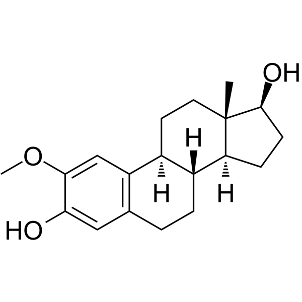
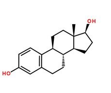
2-Methoxyestradiol Chemical Structure
CAS No. : 362-07-2
| 规格 | 价格 | 是否有货 | 数量 |
|---|---|---|---|
| 10 mM * 1 mL in DMSO | ¥500 | In-stock | |
| 10 mg | ¥400 | In-stock | |
| 50 mg | ¥1000 | In-stock | |
| 100 mg | ¥1700 | In-stock | |
| 500 mg | ¥5100 | In-stock | |
| 1 g | 询价 | ||
| 5 g | 询价 |
* Please select Quantity before adding items.
2-Methoxyestradiol 相关产品
•相关化合物库:
- Natural Product Library Plus
- Drug Repurposing Compound Library Plus
- Clinical Compound Library Plus
- Bioactive Compound Library Plus
- Apoptosis Compound Library
- Cell Cycle/DNA Damage Compound Library
- Immunology/Inflammation Compound Library
- Metabolism/Protease Compound Library
- NF-κB Signaling Compound Library
- Stem Cell Signaling Compound Library
- Natural Product Library
- Anti-Cancer Compound Library
- Clinical Compound Library
- Autophagy Compound Library
- Human Endogenous Metabolite Compound Library
- Anti-Aging Compound Library
- Drug Repurposing Compound Library
- Antioxidants Compound Library
- Lipid Compound Library
- Oxygen Sensing Compound Library
- Ferroptosis Compound Library
- Phenols Library
- Pyroptosis Compound Library
- Cytoskeleton Compound Library
- Orally Active Compound Library
- FDA Approved & Pharmacopeial Drug Library
- Anti-Lung Cancer Compound Library
- Mitochondria-Targeted Compound Library
- Food-Sourced Compound Library
- Rare Diseases Drug Library
| 生物活性 |
2-Methoxyestradiol (2-ME2), an orally active endogenous metabolite of 17β-estradiol (E2), is an apoptosis inducer and an angiogenesis inhibitor with potent antineoplastic activity. 2-Methoxyestradiol also destablize microtubules. 2-Methoxyestradio, also a potent superoxide dismutase (SOD) inhibitor and a ROS-generating agent, induces autophagy in the transformed cell line HEK293 and the cancer cell lines U87 and HeLa[1][2][3][4][5][6]. |
||||||||||||||||
|---|---|---|---|---|---|---|---|---|---|---|---|---|---|---|---|---|---|
| IC50 & Target |
|
||||||||||||||||
| 体外研究 (In Vitro) |
2-Methoxyestradiol (2-ME) (5-100 μM) inhibits assembly of purified tubulin in a concentration-dependent manner, with maximal inhibition (60%) at 200 μM 2-Methoxyestradiol (2ME2). In living interphase MCF7 cells at the IC50 for mitotic arrest (1.2 μM), 2-Methoxyestradiol significantly suppresses the mean microtubule growth rate, duration and length, and the overall dynamicity, consistent with its effects in vitro, and without any observable depolymerization of microtubules. 2-Methoxyestradiol induces G2-M arrest and apoptosis in many actively dividing cell types while sparing quiescent cells. 2-Methoxyestradiol binds to tubulin at or near the colchicine site, it inhibits microtubule assembly, and high concentrations have been shown to depolymerize microtubules in cells[1]. 上海金畔生物科技有限公司 has not independently confirmed the accuracy of these methods. They are for reference only. |
||||||||||||||||
| 体内研究 (In Vivo) |
To investigate the effect of 2-Methoxyestradiol (2-ME2) on uveitis development, C57BL/6 mice are randomly assigned into two groups and immunized with IRBP peptide. 2ME2 group starts 2-Methoxyestradiol (15 mg/kg) intraperitoneally from day 0 to day 13 while control group is given with vehicle. The disease score of 2-Methoxyestradiol (2ME2) group is 0.30±0.30, significantly lower than that of control group 2.09±0.28 (p<0.05), each group containing 5 mice[3]. 上海金畔生物科技有限公司 has not independently confirmed the accuracy of these methods. They are for reference only. |
||||||||||||||||
| Clinical Trial |
|
||||||||||||||||
| 分子量 |
302.41 |
||||||||||||||||
| Formula |
C19H26O3 |
||||||||||||||||
| CAS 号 |
362-07-2 |
||||||||||||||||
| 中文名称 |
二甲氧基雌二醇;二甲氧雌二醇 |
||||||||||||||||
| 运输条件 |
Room temperature in continental US; may vary elsewhere. |
||||||||||||||||
| 储存方式 |
|
||||||||||||||||
| 溶解性数据 |
In Vitro:
DMSO : ≥ 100 mg/mL (330.68 mM) H2O : < 0.1 mg/mL (insoluble) * “≥” means soluble, but saturation unknown. 配制储备液
*
请根据产品在不同溶剂中的溶解度选择合适的溶剂配制储备液;一旦配成溶液,请分装保存,避免反复冻融造成的产品失效。 In Vivo:
请根据您的实验动物和给药方式选择适当的溶解方案。以下溶解方案都请先按照 In Vitro 方式配制澄清的储备液,再依次添加助溶剂: ——为保证实验结果的可靠性,澄清的储备液可以根据储存条件,适当保存;体内实验的工作液,建议您现用现配,当天使用; 以下溶剂前显示的百
|
||||||||||||||||
| 参考文献 |
|
| Kinase Assay [1] |
Microtubule protein (2.75 mg/mL) is assembled to steady-state [in 100 mM PIPES containing 1 mM EGTA and 1 mM MgSO4 (PEM100) and 1 mM GTP, 35°C for 45 minutes] containing 2-Methoxyestradiol (final drug concentrations of 1-500 μM). Final DMSO and ethanol concentrations are adjusted to 1% and 5%, respectively. Concentrations of 2-Methoxyestradiol ≤ 5 μM have no effect on microtubule polymer mass, and thus 20 to 500 μM 2-Methoxyestradiol is used for most of the experiments. Incubation with 2-Methoxyestradiol is carried out for 30 minutes, at which time microtubule depolymerization is maximal, and microtubules are centrifuged at 35°C for 30 minutes and the supernatant is removed from the pellets. Microtubule pellets are solubilized overnight in 0.2 M NaOH and the protein concentrations of supernatants and pellets are determined[1]. 上海金畔生物科技有限公司 has not independently confirmed the accuracy of these methods. They are for reference only. |
|---|---|
| Cell Assay [1] |
MCF7 breast carcinoma cells stably transfected with green fluorescent protein (GFP)-α-tubulin are cultured in DMEM supplemented with nonessential amino acids, 0.1% penicillin/streptomycin, 10% fetal bovine serum, and 0.4 mg/mL G418 at 37°C in 5% CO2. Transfection of MCF7 cells with GFP-α-tubulin is carried out. To evaluate mitotic indices, cells are plated at a concentration of 6×10⁴/2 mL into six-well plates. After 48 hours, cells are incubated in the absence or presence of 2-Methoxyestradiol at concentrations ranging from 100 nM to 30 μM for 20 hours. To collect both floating and attached cells, medium is collected; attached cells are rinsed with Versene (137 mM NaCl, 2.7 mM KCl, 1.5 mM KH2PO4, 8.1 mM Na2HPO4, and 0.5 mM EDTA), detached by trypsinization, and added back to the medium. Cells are collected by centrifugation and fixed with 10% formalin for 30 minutes, permeabilized in ice-cold methanol for 10 minutes, and stained with 4′,6-diamidino-2-phenylindole to visualize nuclei. Results are the mean and SE of seven experiments in each of which 500 cells are counted for each concentration. The mitotic IC50 is the drug concentration that induced one half of the maximal mitotic accumulation[1]. 上海金畔生物科技有限公司 has not independently confirmed the accuracy of these methods. They are for reference only. |
| Animal Administration [3][4] |
Mice[3] 上海金畔生物科技有限公司 has not independently confirmed the accuracy of these methods. They are for reference only. |
| 参考文献 |
|
所有产品仅用作科学研究或药证申报,我们不为任何个人用途提供产品和服务

