上海金畔生物科技有限公司为生命科学和医药研发人员提供生物活性分子抑制剂、激动剂、特异性抑制剂、化合物库、重组蛋白,专注于信号通路和疾病研究领域。
PAC-1 (Synonyms: Procaspase activating compound 1) 纯度: 99.93%
PAC-1 是一种 procaspase-3 激活剂,诱导癌细胞凋亡,EC50 为 2.08 μM。
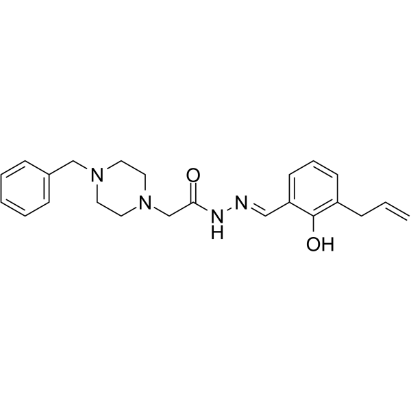
PAC-1 Chemical Structure
CAS No. : 315183-21-2
| 规格 | 价格 | 是否有货 | 数量 |
|---|---|---|---|
| 10 mM * 1 mL in DMSO | ¥550 | In-stock | |
| 10 mg | ¥500 | In-stock | |
| 50 mg | ¥1400 | In-stock | |
| 100 mg | ¥2500 | In-stock | |
| 500 mg | ¥10000 | In-stock | |
| 1 g | 询价 | ||
| 5 g | 询价 |
* Please select Quantity before adding items.
PAC-1 相关产品
•相关化合物库:
- Drug Repurposing Compound Library Plus
- Clinical Compound Library Plus
- Bioactive Compound Library Plus
- Apoptosis Compound Library
- Anti-Cancer Compound Library
- Clinical Compound Library
- Autophagy Compound Library
- Drug Repurposing Compound Library
- Pyroptosis Compound Library
- Anti-Blood Cancer Compound Library
- Targeted Diversity Library
| 生物活性 |
PAC-1 is a procaspase-3 activator that induces apoptosis in cancer cells with an EC50 of 2.08 μM. |
||||||||||||||||
|---|---|---|---|---|---|---|---|---|---|---|---|---|---|---|---|---|---|
| IC50 & Target[1] |
|
||||||||||||||||
| 体外研究 (In Vitro) |
PAC-1 activates procaspase-3 with an EC50 of 2.08 μM. PAC-1 exhibits an enhanced zinc chelating ability (EC50= 7.08 μM). PAC-1 induces leukemia cell death with IC50 of 4.03 μM, which is consistent with the values reported by other investigators. PAC-1 treatment also results in death of other malignant cells in a concentration-dependent manner with IC50s ranging from 4.03 to 53.44μM. The overall mean IC50 in the fifteen malignant cell lines is 0.88 mM for WF-210 and 19.40 μM for PAC-1. In contrast, the sensitivity of the normal human cells (PBL, L-02, HUVEC and MCF 10A) to WF-210 is 2.6-fold lower (mean IC50=412.34 μM) than PAC-1 (mean IC50=158.29 μM)[1]. Procaspase-activating compound-1 (PAC-1) is the first direct caspase-activating compound discovered. PAC-1 treatment upregulates Ero1α in multiple cell lines, whereas silencing of Ero1α significantly inhibits calcium release from ER and cell death[2]. 上海金畔生物科技有限公司 has not independently confirmed the accuracy of these methods. They are for reference only. |
||||||||||||||||
| 体内研究 (In Vivo) |
To evaluate the in vivo effect of WF-210 on the growth of malignant tumors, we examined the ability of WF-210 to suppress tumor growth in mouse Hep3B and MDA-MB-435 xenograft models. These two cell lines express procaspase-3 at relatively high levels. Tumors induced by xenografts of the liver cancer cell Hep3B are allowed to develop and grow to a size of 100 mm3, after which WF-210 (2.5 mg/kg) or PAC-1 (5.0 mg/kg) is given daily for two weeks by intravenous (i.v.) administration. As shown in both PAC-1 and WF-210 significantly inhibits the growth of Hep3B tumor xenografts[1]. 上海金畔生物科技有限公司 has not independently confirmed the accuracy of these methods. They are for reference only. |
||||||||||||||||
| Clinical Trial |
|
||||||||||||||||
| 分子量 |
392.49 |
||||||||||||||||
| Formula |
C23H28N4O2 |
||||||||||||||||
| CAS 号 |
315183-21-2 |
||||||||||||||||
| 中文名称 |
半胱天冬酶原活化物1 |
||||||||||||||||
| 运输条件 |
Room temperature in continental US; may vary elsewhere. |
||||||||||||||||
| 储存方式 |
|
||||||||||||||||
| 溶解性数据 |
In Vitro:
DMSO : 50 mg/mL (127.39 mM; Need ultrasonic) H2O : < 0.1 mg/mL (insoluble) 配制储备液
*
请根据产品在不同溶剂中的溶解度选择合适的溶剂配制储备液;一旦配成溶液,请分装保存,避免反复冻融造成的产品失效。 In Vivo:
请根据您的实验动物和给药方式选择适当的溶解方案。以下溶解方案都请先按照 In Vitro 方式配制澄清的储备液,再依次添加助溶剂: ——为保证实验结果的可靠性,澄清的储备液可以根据储存条件,适当保存;体内实验的工作液,建议您现用现配,当天使用; 以下溶剂前显示的百
|
||||||||||||||||
| 参考文献 |
|
| Kinase Assay [1] |
Various concentrations of WF-210 or PAC-1 are added to procaspase-3 in buffer containing 50 mM HEPES, 0.1% CHAPS, 10% glycerol, 100 mM NaCl, 0.1 mM EDTA, 10 mM DTT pH 7.4,and incubated for 12 h at 37°C. The final volume is 10 mL and the final concentration of procaspase-3 is 1 mM. Then 40 mL of the substrate Ac-DEVD-pNA (final concentration 0.4 mM) in buffer containing 50 mM HEPES pH 7.4, 100 mM NaCl, 10 mM DTT, 0.1 mM EDTA disodium salt, 0.10% CHAPS, 10% glycerol is added and the absorbance of the plate is read at 405 nm for a total of 1 h. The slope of the linear portion for each well is determined as the enzyme activity[1]. 上海金畔生物科技有限公司 has not independently confirmed the accuracy of these methods. They are for reference only. |
|---|---|
| Cell Assay [1] |
Cell viability is measured using the MTT method or the Cell Titer-Glo luminescent assay. For the MTT assay, the cells (1×10⁵ cells/mL) are seeded into 96- well culture plates. After overnight incubation, cells are treated with various concentrations of agents (PAC-1, WF-210 or other agents) for 24 or 72 h. Then 10 mL MTT solution (2.5 mg/mL in PBS) is added to each well, and the plates are incubated for an additional 4 h at 37°C. After centrifugation (2500 rpm, 10min), the medium containing MTT is aspirated, and100mL DMSO is added. The optical density of each well is measured at 570 nm with a Biotek Synergy HT Reader. The Cell Titer-Glo kit is used to determine the relative levels of intracellular ATP as a biomarker for live cells[1]. 上海金畔生物科技有限公司 has not independently confirmed the accuracy of these methods. They are for reference only. |
| Animal Administration [1] |
Mice[1] 上海金畔生物科技有限公司 has not independently confirmed the accuracy of these methods. They are for reference only. |
| 参考文献 |
|
所有产品仅用作科学研究或药证申报,我们不为任何个人用途提供产品和服务

