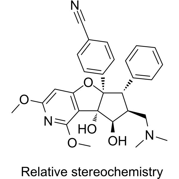
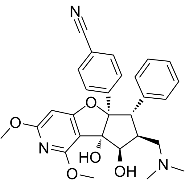
体外研究
(In Vitro) |
Zotatifin induces the formation of a stable ternary complex [eIF4A-RNA-eFT226]. Zotatifin increases the residence time for eIF4A1 binds to an AGAGAG RNA surface, the Kd values are 0.021 μM and 8.0 μM, respectively for eFT226 presence or absence[1].Zotatifin inhibits in vitro translation as a sequence-dependent manner, the IC50 values are 1.5 nM, 13.8 nM, 92.5 nM, and 217.5 nM, respectively in an MDA-MB-231 cell line with transiently transfected AGAGAG, GGCGGC, CCGCCG and CAACAA 5’-UTRs-containing sequences[1].Zotatifin (0.0001 μM-1 μM; 72 hours) inhibits tumor cells growth as a dose-dependent manner. It shows a potent anti-proliferative activity (GI50<15 nm) in mda-mb-231 tumor cells, but eif4a1 f163l mutation rescues eft226 anti-proliferative activity[1].Zotatifin (0.0001 μM-1 μM; 72 hours) inhibits tumor cell growth, exhibits GI50 values for TMD8, SU-DHL-2, HBL1, Pfeiffer, SU-DHL-6, SU-DHL-10, VAL, Carnaval, U2973, Ramos, Jeko1, Mino, and Rec-1 cells are 4.1 nM, 3 nM, 5.6 nM, 3.7 nM, 5.3 nM, 7.3 nM, 6.6 nM, 4.4 nM, 4.2 nM, 4.6 nM, 7.9 nM, 11.2 nM and 11.8 nM, respectively[1].Zotatifin (30 μM-100 μM; 3 or 24 hours) results in translational regulation of oncogenic protein, decreases MYC,CCND3,BCL2 and MCL1 protein expression as a time- and dose-dependent manner[1].The anti-viral activity of Zotatifin is demonstrated by various assays: such as TCID50 assay, Plaque assay, NP-staining assay, et al[2].Zotatifin (10 nM, 100 nM, 200 nM, 500 nM, 2 μM, 10 μM; 1 or 2 hours pre-treatment before virus isolates) decreases the detection of the viral NP protein and reduces viral infectivity in a concentration-dependent matter in Vero E6 cells cells infected with SARS-CoV-2 isolates[2].
上海金畔生物科技有限公司 has not independently confirmed the accuracy of these methods. They are for reference only.
Cell Viability Assay[1]
| Cell Line: |
MDA-MB-231 tumor cells |
| Concentration: |
0.0001 μM, 0.001 μM, 0.01 μM, 0.1 μM, 1 μM |
| Incubation Time: |
72 hours |
| Result: |
Inhibited cell growth with a GI50 of 15 nM, and F163L mutant rescued anti-proliferative effects. |
Cell Proliferation Assay[1]
| Cell Line: |
DLBCL-ABC; DLBCL-GCB; Burkitt; and MCL tumor type cells |
| Concentration: |
0.0001 μM, 0.001 μM, 0.01 μM, 0.1 μM, 1 μM |
| Incubation Time: |
72 hours |
| Result: |
Inhibited cell growth with GI50 values ranging from 3 nM to 20 nM. |
Cell Proliferation Assay[1]
| Cell Line: |
TMD8 and Pfeiffer DLBCL tumor cells |
| Concentration: |
30 μM; 100 μM |
| Incubation Time: |
3 or 24 hours |
| Result: |
Decreased MYC, CCND3, Bcl2,and MCL1 protein levels. |
|
体内研究
(In Vivo) |
Zotatifin (intravenous injection; 1 mg/kg; 14-22 days) decreases tumor volume, inhibits the TMD8 xenograft-bearing, HBL1 xenograft-bearing, Pfeiffer xenograft-bearing, SU-DHL-6 xenograft-bearing, SU-DHL-10 xenograft-bearing and Ramos-bearing animals’tumor growth as percentage of 97%, 87%, 70%, 83%, 37% and 75%, respectively[1].Zotatifin (intravenous injection; 0.001 mg/kg-1 mg/kg; 15 days) inhibits the growth of B-cell lymphoma xenografts and is well-tolerated against B-cell lymphoma xenograft models in vivo[1].
上海金畔生物科技有限公司 has not independently confirmed the accuracy of these methods. They are for reference only.
| Animal Model: |
B-cell lymphoma xenograft model[1] |
| Dosage: |
0.001 mg/kg; 0.1 mg/kg; 1 mg/kg |
| Administration: |
Intravenous injection; 15 days |
| Result: |
Showed efficacy in B-cell lymphoma xenograft models. |
|


